Introduction
Eczema is a clinical and histological pattern of inflammation of the skin seen in a variety of dermatoses with widely diverse aetiologies. Clinically, eczematous dermatoses are characterized by variable intensity of itching and soreness, and in variable degrees, a range of signs including dryness, erythema, excoriation, exudation, fissu ring, hyperkeratosis, lichenification, papulation, scaling and vesiculation. Histologically, the clinical signs are reflected by a range of epidermal changes including spongiosis (epidermal oedema) with varying degrees of acanthosis and hyperkeratosis, accompanied by a lymphohistiocytic infiltrate in the dermis.1 Their pattern and prevalence however vary between countries and various regions of the same country due to varying ecological factors.
Some forms of eczemas are altered by regional variation in structure and function of the skin, and these may modify its appearance in regions such as hands and lower legs.2
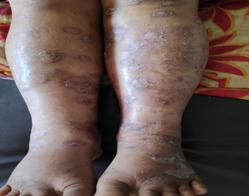
Legs and feet are affected by various eczemas. Classically stasis dermatitis affects lower legs [Figure 1]. Other eczemas more likely to occur are nummular eczema, juvenile plantar dermatosis, pompholyx, lichen simplex chronicus and allergic contact dermatitis. They are usually chronic, recurrent and difficult to control. They may make patients unable to perform daily activities because of pain caused by fissures or because of secondary infections. They may prevent the wearing of shoes, especially when shoes are the cause of the dermatosis.2
Good data is available regarding the patterns of hand dermatitis but there is hardly any study on the patterns of lower leg and feet eczemas. Hence, we undertook this study to determine frequency and patterns of lower leg and foot eczema.
Materials and Methods
A descriptive study of lower leg and foot eczema was carried out among the patients from July 2018 to June 2019 in Department of Dermatology, Silchar Medical College, Assam. The study was conducted after getting clearance from Institutional Ethical Committee.
Diagnosis of eczemas involving lower leg and foot was made utilizing standard definitions accepted in the literature.1,3,4,5,6
Associated symptoms such as pain, pruritus, dryness, scaling, redness, crusting, fissuring and oozing were observed. An occupational history with emphasis into the common allergens that the patient may be exposed to was elicited followed by a detailed clinical examination and all the findings were recorded on a proforma.
After analyzing the history and clinical examination findings, type of eczema the patient was suffering from could be ascertained. Patients were prescribed appropriate medications and advised to visit after the lesions subsided. The data was tabulated and analyzed.
Results
One hundred patients with eczema of lower leg and foot were included in this study. Mean age of 100 patients in this study was 40 years with 52(52%) male and 48(48%) were females with male to female ratio 1.08:1.
The most common age group affected was 31-45 years (35 cases, 35%) followed by 46-60 years age group (25 cases,25%), 16-30 years (17 cases, 17%), less than or equal to 15 years (10cases, 10%), 61-75 years (8 cases, 8%) and more than 75 years age group (5 cases,5%). Majority of the male (52 cases, 52%) were in 31-45 and 46-60 year age groups; whereas majority of the females (48 cases, 48%) were in 31-45 years age group [Table 1]. Majority of the study population (40 cases, 40%) were farmers, followed by housewives (27cases, 27%), students (10 cases, 10%), daily wage laborers (18 cases, 18%), office workers (3 cases, 3%), and others (2,2%cases).
Table 1
| Age group(years) | Total cases | Male | Female |
| 0-15 | 10 | 3 | 7 |
| 16-30 | 17 | 10 | 7 |
| 31-45 | 35 | 22 | 13 |
| 46-60 | 25 | 13 | 12 |
| 61-75 | 8 | 2 | 6 |
| >75 | 5 | 2 | 3 |
| Total | 100 | 52 | 48 |
Mean duration of eczema was 33 months and the range was 5 days to 24 years. Eczemas were bilateral in 75 cases (75%). Rest were unilateral.
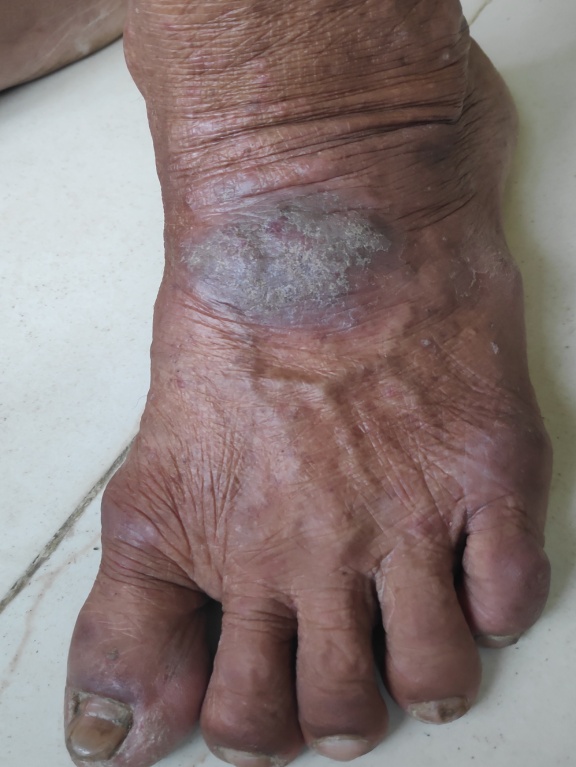
Sites of involvement in decreasing order of frequency were as follows: dorsum of foot (41 cases, 41%)[Fig 6], lateral aspect of lower leg (4 cases, 4%), medial aspect of lower leg (5 cases,5%), ankle (15 cases, 15 %), dorsum of toes (5 cases, 5%), instep of sole (3 cases, 3%), anterior part of sole (3 cases, 3 %), toe web spaces (4 cases, 4 %), plantar aspect of toes (3cas es, 3%), posterior aspect of foot (4 cases, 4%), posterior part of sole (8cases, 8 %) and posterior aspect of leg (5 case, 5%)[Table 2].
Table 2
Table 3
A total of 14 types of lower leg and foot eczema were noted in this study.
Correlation with clinical parameters
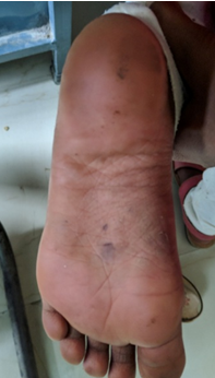
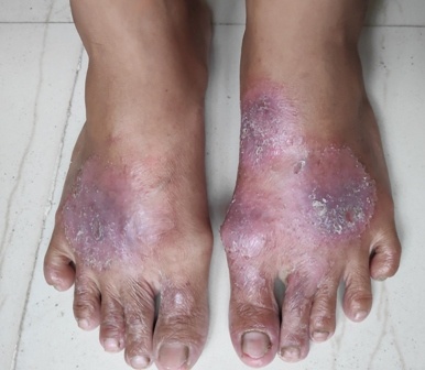
Age and type of eczema: The occurrence of the type of eczema was found to be influenced by age. On analysis, the most common eczema in patients younger than 15 years of age was juvenile plantar dermatosis (5 cases, 50%)[fig 2]. The most common eczema in 16-30 year age group was cumulative irritant contact dermatitis (10 cases, 58.8%), followed by discoid eczema (5 cases,29.4%) and stasis dermatitis (2 cases,11.7%). The most common eczema in 31-45 year age group was lichen simplex chronicus (20 cas es, 57.1%) followed by atopic dermatitis (7 cases, 20%) and pompholyx (6 cases, 17.1%). The most common eczema in 46-60 year age group was lichen simplex chronicus (10 cases, 40%)[Figure 4], followed by hyperkeratotic eczema (6 cases, 24%) and infected eczema (6 cases,24%)[Figure 3].The most common eczema in 61-75 year age group was asteatotic eczema (7 cases,87.5% cases).
Gender and type of eczema: Gender had no bearing on the occurrence of lichen simplex chronicus and discoid eczema [Fig. 5]. However some eczemas such as cumulative irritant contact dermatitis and pompholyx were observed more in female patients. Stasis eczema was also more frequent in females. Asteatotic eczema were relatively infrequent in female patients.
Occupation and type of eczema: Infected eczema was found to be common in farmers (5 out of 7 cases) and cumulative irritant contact dermatitis in housewives (6 out of 10 cases). The most common types of eczema in students were juvenile plantar dermatosis (4 out of 5 cases) and allergic contact dermatitis (4 out of 6 cases). In daily wage workers lichen simplex chronicus (14 out of 30 cases) was the most common eczema. In office workers, the most common eczema found was lichen simplex chronicus (9 out of 30 cases).
Duration and type of eczema: In most cases of allergic contact dermatitis (4 out of 6 cases), infected eczema (6 out of 7 cases) and pompholyx (5 out of 7 cases), stasis dermatitis (2 out of 4 cases), the duration of eczema was 6 months or less. Most cases of lichen simplex chronicus (25 out of 30 cases), discoid eczema (4 out of 5 cases), cumulative irritant contact dermatitis (8 out of 10 cases), juvenile planar dermatosis (5 out of 5 cases), hyperkeratotic eczema (4 out of 6 cases) and asteatotic eczema (5 out of 7 cases) had their disease for 6 months to 5 years. 2 cases of lichen simplex chronicus and 1 cases of hyperkeratotic plantar eczema had their disease duration 25 years.
Distribution and type of eczema: Although unilateral distribution was sometimes seen, the following types of eczemas were predominantly bilateral: lichen simplex chronicus, podopompholyx, discoid eczema, allergic contact dermatitis, stasis eczema, hyperkeratotic plantar eczema. Unilateral distribution was seen in the following types of eczema: some cases of lichen simplex chronicus, infected eczema, post traumatic eczema and dermatophytide.
Site of involvement and type of eczema: Juvenile plantar dermatosis, hyperkeratotic eczema, pompholyx and asteatotic eczemas were diagnosed because of their distinctive morphology and sites of involvement. Dorsum of foot was involved in lichen simplex chronicus (20 cases out of 30), discoid eczema (2 cases out of 5 cases), allergic contact dermatitis (3 cases out of 6 cases) and stasis eczema (2 cades out of 4 cases). Lateral aspect of leg was involved in lichen simplex chronicus (3 cases out of 30 cases), discoid eczema (1 cases out of 5). Medial aspect of leg was involved in 2 out of 2 cases of stasis eczema. Ankle was involved in LSC in 15 cases out of 30 cases of LSC. Instep of sole was involved in 1 out of 2 cases of infective eczema.
Discussion
Hanifin et al. reported most frequent ages at onset for empirically defined eczema in a population based survey of eczema in the range of 18 to 29 years 7 The age of patients in present study ranged from 0- 80 years with a mean of 40 years. Most patients belonged to 31-45 year age group.
Lichen simplex chronicus (30%) was the predominant group of lower leg and foot eczema which is consistent with another study done by Abhijit Chougule, Devinder Mohan Thappa in which it’s incidence was 36%2 Lichen simplex chronicus may be primary (arising on tissue with a normal appearance) or secondary (superimposed on other underlying disease). It occurs most commonly between the ages of 30-50 years.1 Almost any area may be affected by LSC, but most common sites are those that are conveniently reached. It can be either unilateral or bilateral reflecting a preference for scratc hing with the dominant hand.1
Stasis eczema typically affects middle-aged or elderly, especially female, patients and rarely occurs before the fifth decade of life, except in patients with acquired venous insufficiency. In a study of incidence of various types of eczemas over 27 years in Ipswich, UK, gravitational eczema was recorded in 4% cases.1 In our study, stasis eczema was observed in 4 cases (4%) out of a total 100 cases of lower leg and foot eczema, with a mean age of 55.6 years. This eczema was seen most commonly in farmers (3 cases) as they are involved in standing as well as mobile field work. In a study conducted by Abhijit Chougule, Devinder Mohan Thappa in which it’s incidence was 7.5%2 In another study done by S. Vijay Shankar, V. N. S. Ahamed Shariff, S. Nirmala, the prevalence is found to be highest in the age group of 50 to 60 years (42.5%).8 The prevalence of the disease was common in the male population which is also found in our study.
Jones et al reported 102 cases of juvenile plantar dermatosis, mean age at onset of disease was 7.3 years and the mean duration of disease was 8.4 years.9 In our study, juvenile plant ar dermatosis was observed in 5 cases (5%) and age of all patients were below 15 years and the findings were in concurrence with the literature.
Cumulative irritant contact dermatitis develops as a consequence of multiple incidents of sub threshold damage to the skin and was a unique feature of our study. It was observed in 10 cases (10 %), with a mean age of 25 years. This eczema was observed most commonly in housewives (6 cases), probably because many Indian housewives are involved with household work including washing clothes and utensils with detergents and water. All 10 cases had bilateral eczema, involving most commonly insteps of soles and toe web spaces followed by margins of feet, and dorsal aspect of toes. In a study conducted by Abhijit Chougule, Devinder Mohan Thappa in which it’s incidence was 5% with a mean age of 28.3 years.2
Hyperkeratotic eczema occurs commonly in middle-aged to elderly men and is normally very refractory to treatment whereas asteatotic eczema commonly occurs on the shins of elderly patients.10 Clinically, there are several forms of asteatotic eczema which are either localized or generalized and which have different implications.11 Asteatotic eczema of legs was observed in 7 cases (7%) in our study. In a study done by Iqbal T, Kapadia N, Athar S, Iqbal S, Shahmoona S, Mansoor M the incidence of asteatotic eczema was found to be 10%.12
Infective eczema is an eczema that is caused by microorganisms or their products, and which clears when organisms are eradicated. It must be distinguished from infected eczema in which eczema resulting from some other cause is complicated by secondary bacterial or viral invasion of the broken skin. These two conditions may coexist and distinction can be difficult.1 In the present study, infected eczema affecting lower legs and feet were observed in 7 cases (7%) while infective eczema was observed in two cases (2%) only. In a study done by Abhijit Chougule, Devinder Mohan Thappa, it’s incidence was found 5% and 1% respectively.2
Allergic contact dermatitis of legs and feet is mainly associated with the use of footwear and topical medicaments for eczema.13,14 In the present study, allergic contact dermatitis was suspected in 6 cases (6%). It occurs only in sensitized individuals and population estimates vary from 1.7% to 6%.15 The lower leg is particularly prone to contact allergy. ACD from medicaments and dressings predominates, especially in those with varicose eczema and ulcer. The feet have a peculiar anatomical feature, such as the highest concentration of eccrine sweat glands in the plantar region; this leads to increased maceration and enhances the absorption of chemicals, potentially increasing the risk of ACD. Common allergens implicated are potassium dichromate present in footwear, nickel sulfate, mercapto mix, and mercaptobenzothiazole. Potassium dichromate and nickel were the common allergens reported in males and females respectively. Neomycin was the common medication responsible for dermatitis medicamentosa.15
Conclusion
Lower leg and foot eczemas are one among the many common dermatological disorders that are observed in a dermatology out-patients department. In our study, lichen simplex chronicus is found to be the most common eczema affecting lower limb. There is significant association between the various risk factors and development of eczema. This study confirms the importance of age, sex, occupation, environmental factors etc. in development of eczema.A wide range of presentation was noticed in our study. It is important to identify the high risk patients at an early stage and appropriate preventive measures to be initiated to prevent complications.


