Introduction
Elastosis perforans serpiginosa is rare perforating dermatoses in which there is transepidermal elimination of elastic fibres from papillary dermis into the epidermis. In year 1955 scientist Miescher explained that extruded out is elastin and suggested the term elastomer intrapaillare perforans veruciforme, which was later termed as EPS by Dammert and Putkonem.1, 2 It usually present in early period of life in between 5 to 20 years of age group with males predominantly affected. In 40% of cases of EPS are generally associated with many heritable connective tissue diseases like Pseudoxanthoma Elasticum, Marfan syndrome, Osteogenesis Imperfecta, Acrogeria, Ehler Danlos Syndrome and Down syndrome patients.3 Penicillamine induced EPS is seen in which overproduction of elastic fibres is seen. On histopathology there is acanthosis and hyperkeratoses is seen. There is increase in amount of elastic fibres in papillary dermis and inflammation along with transepidermal elimination of elastic fibres is present.4 Condition is generally self limiting with no proven benefit.5
Case Report
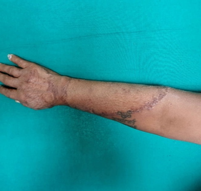
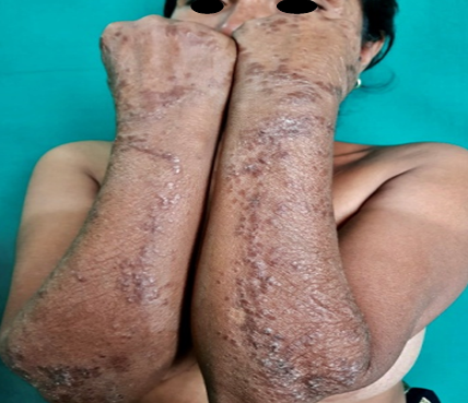
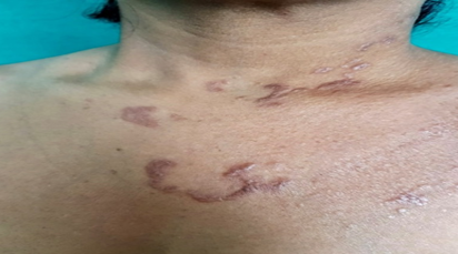
A 45 year old diabetic female presented in OPD with chief complaints of asymptomatic dark coloured raised lesions starting from face and sides of neck and gradually progressing to involve chest and flexor aspect of upper extremities in bilaterally symmetrical pattern for last 4 years. On cutaneous examination multiple hyperpigmented keratotic papular lesions coalescing to form plaques were arranged in annular, serpiginous and polycyclic pattern over face, sides of neck, chest and forearms. Lesions present over forearms had largest diameter (15-20 cm), distributed in bilateral symmetrical pattern with obvious central sparing and atrophy. Margins were well delineated with no associated symptoms. Few individual had an impression of central keratotic core. [Figure 1, Figure 2, Figure 3]
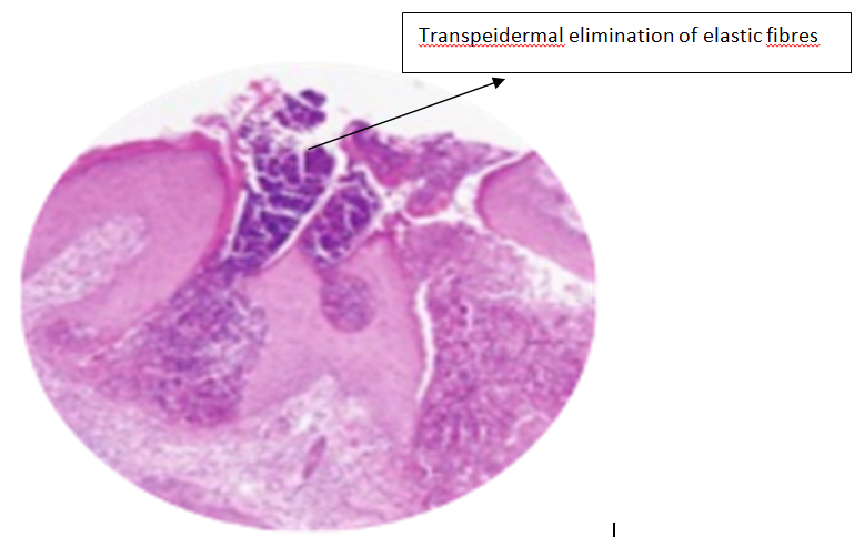
There was no family history of similar disease. Family, past and drug history were non supportive. Patient underwent clinical and systemic examination followed by routine and histopathological investigations. Random blood sugar were controlled on anti diabetic medication. On histopatholgy hyperkeratotic and acanthotic epidermis with transepidermal channel having nuclear debris and abnormal elastic fibres. [Figure 4].
Patient was explained about the course and prognosis of the disease and was started on oral and topical retinoids. Later on she was lost to follow up.
Multiple hyperpigmented keratotic papular lesions coalesce to form plaque in annular, serpigimous and polycyclic pattern present over forearms with central sparing and atrophy.
Discussion
EPS is one of the four primary perforating disorder. It is more common in male with M:F ratio 4:1. Most common age group affected was 5 to 20 years. Etiopathogenesis is unclear in 60% cases while in rest it was associated with Down’s syndrome and other hereditary connective tissue disorder like Ehler Danlos syndrome and Marfan syndrome.3 There is primary defect in the dermal elastin which induces the extrusion of elastic fibres abnormally. The lesions are more common on those areas were there is more wear and tear. Penicillamine is one drug which is responsible for EPS in which histopathological appearance is like ‘bramble bush’ or ‘lumpy-bumpy’.6 Main stay of diagnosis is clinical appearance and histopathology. The disease is generally self limiting. Successful treatment with tazarotene 0.1% gel was presented by Outland et al and Kelly and Purcell noticed that reduction of skin lesions with imiquimod for a period of 10 weeks.7, 8


