Case Report
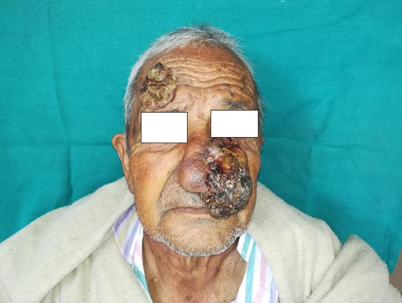
81 year old male presented with two painless fungating growths on face for last 5 months. It started as a nodule which progressed to massive growth, one on left lateral nasal wall and other on right side of forehead after a period of 2months. There was foul smelling discharge and occasional bleeding from the lesions. Patient complained of difficulty in breathing from left lateral nostril for last 1 month.
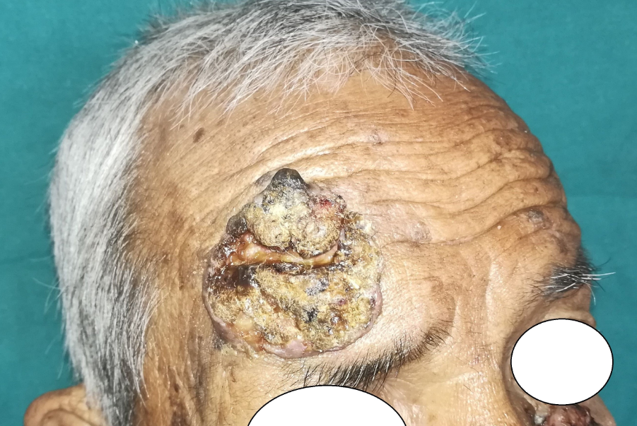
On local examination there was a giant exophytic growth arising from left lateral nasal wall of size approximately 10cm*6cm*5cm, encroaching whole left lateral nasal wall, left upper-lower lip and left cheek. Its surface was irregular and contained patches of necrotic tissue at places (Figure 1). Foul smelling seropurulent discharge was also present. The growth was non tender, friable, fixed to underlying nasal cartilages but not breaching the nasal mucosa. Left nostril was collapsed. Another similar growth of size 5cm*5cm*2cm was present on right side of forehead which was not fixed to underlying bone (Figure 2). There was no lymphadenopathy. Systemic examination was within normal limits.
Laboratory tests like complete blood count, liver function test, renal function tests were within normal limits. Computed tomography revealed nasal growth infiltrating left alar and left upper and lower lateral nasal cartilages and just abutting the nasal septum with collapse of left nostril. Forehead growth was involving only skin and subcutaneous tissue. There was no evidence of lymphadenopathy.
Wide local excision was performed under general anaesthesia. Forehead defect was covered with split skin graft taken from right thigh and nasal defect was covered with left paramedian forehead flap (Figure 3).
Histopathological examination of the growth showed moderately differentiated squamous cell carcinoma and all resection margins were free.
Post-operative course was uneventful.
Discussion
Cutaneous SCC is the second most common form of non-melanoma skin cancer, basal cell carcinoma being the first.1 Mostly SCC occurs in photoexposed sites but it has been reported in covered sites as well particularly in dark skinned individuals.2 Persistent sun exposure is the most important risk factor particularly the wavelengths of UVB spectrum.3 Radiation therapy, arsenic exposure etc are the other important etiological agents. Incidence of SCC increases with age but it has also been reported in young though rarely 4
SCC is also common in pre-existing dermatological conditions like actinic keratosis, discoid lupus erythematosus, lupus vulgaris, bowenoid papulosis etc.5
Various treatment modalities include surgical excision, radiotherapy, curettage and electrodessication.5 Surgical resection and reconstruction is most effective mode of treatment which also gives a good cosmetic result. Choice of treatment in elderly is challenging. Due to associated comorbidities surgical management becomes difficult with advancing age but there was no such underlying systemic illness in our case
Moreover such giant tumors can’t be managed conservatively and thus we opted for surgical excision of the tumour. We would like to highlight that advancing age should not be absolute contraindication for surgery as such people should also have a good quality of life when there is no other comorbidity. Surgical excision of the tumour becomes the most reliable method to prevent the progression, metastasis of the tumour.
Giant dual malignancy of the face corrected with surgical excision in an old male is the highlight of our case. To the best of our knowledge we could not find any previous reports with such giant double malignancy of the face.


