Introduction
Syphilis is a common sexually, blood contracted or congenitally transmitted disease. It can be divided into primary syphilis, secondary syphilis, tertiary syphilis, and neurosyphilis. Its florid presentation renders the name “the great imitator” to it.2 Syphilis is a chronic disease which clinically present with a waxing and waning course. The cutaneous manifestations of secondary syphilis include mainly macular, papular or pustular and also lichenoid, papulopustular, psoriasiform, vesicular or corymbiform lesions.2, 3 Although the clinical features allow the diagnosis of secondary syphilis in most cases, it may be at times difficult to differentiate it from other annular maculo-papular dermatoses with scaling. Histology of secondary syphilitic lesions may show considerable variations, depending on the clinical morphology of eruption. Dermoscopy has been most often used to improve the diagnostic accuracy in the clinical evaluation of pigmented skin lesions but it has also been found useful in the assessment of vascular structures that are not visible to the naked eye. Serologic testing remains gold standard for diagnosis of syphilis and penicillin G is treatment of choice. Here by we report a case of secondary syphilis with hyperpigmented macules over palm and soles with hyperpigmented papules over labia majora
Case Report
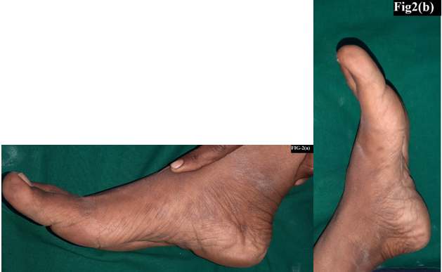
A 23 years old married, nulliparous female presented to dermatology out-patient department with complaint of hyperpigmented lesions over both palms and soles. Cutaneous examination showed multiple ill-defined bilaterally symmetrical, hyperpigmented, discrete round macules of size 5-10 mm over palm and medial border of foot since 2 months. Bilateral inguinal lymph nodes were enlarged, tender, palpable, spherical in shape and with size of 1 cm diameter, soft in consistency, mobile and skin was normal. Bilateral epitrochlear lymph node were palpable. Oro genital examination was normal. Buschke–Ollendorf sign was negative. She had no history of any genital lesions in past. She is married since 7 years and she has history of unprotected penovaginal intercourse with 3 unknown partners before marriage and with multiple partners over a period of last 2 years. There was no significant family history. A 4 mm skin punch biopsy was taken which showed squamous epithelium with mounds of orthokeratosis and mild hypergranulosis. The underlying papillary dermis shows sparse to mild perivascular lymphoplasmacytic inflammatory infiltrate and was conclusive of secondary syphilis. Rapid plasma regain test was positive with 1:32 titer. Patient’s Treponema pallidum hemagglutination assay was positive. Patients husband had no complaints and his RPR & TPHA test was negative. With a diagnosis of palmoplantar syphilis, benzathine penicillin 2.4 MU intramuscular injection was given.
Figure 1
Multiple ill-defined bilaterally symmetrical, hyperpigmented, discrete round macules of size 5-10 mm over both palm

Discussion
Syphilis is a sexually transmitted disease which is caused by Treponema pallidum. The primary lesions mostly occur over the genitals, anus, oral cavity, lips, pharyngeal, and nipple-areola.4 Treponema pallidum is a motile, gram positive bacterium transmitted via sexual, oral-genital or oral-anal contact, contact with contaminated material, and intra-uterine transmission.5
Clinically, syphilis is classified into four stages: Primary, secondary, latent (early and late) and tertiary. The first stage is manifested by painless ulcers which are called hard chancre that disappear within 6 weeks. Syphilis chancre is painless and may be localized in areas out of sights such as rectum and vagina. If it remains untreated, secondary stage appears within 10 weeks which includes low grade fever, malaise, headache, lymphadenopathy and cutaneous eruption which can be macular, papular, pustular etc. Macular syphilide presents as pinkish or coppery red or greyish macules which are discrete, usually less than 1 cm in size, round or oval in shape and non-scaly. Papular syphilide presents as dull red or skin colored or greyish, discrete, annular, circinate or corymbose pattern. Secondary syphilis also characterized by moth eaten alopecia, lichen syphiliticus, mucous patches and snail track ulcers in buccal mucosa also occur. If not recognized and treated properly, syphilis may progress into the devastating tertiary stage, which results in spread of spirochete to nervous system, heart and bone.6 In secondary syphilis Iritis, anterior uveitis, arthritis, and glomerulonephritis or nephrotic syndrome also seen. Circulating immune complexes that contain treponemal antigen and human fibronectin, together with antibody and complement, are present in this stage of infection, and their deposition in relevant organs is thought to play a role in the pathogenesis of these syndromes.7 Usually all patients who have secondary syphilis have antibody to cardiolipin, detectable in tests such as the Rapid Plasma Reagin (RPR) or VDRL tests.8 Antibody to treponemal outer membrane proteins, as measured in tests such as the MHA-TP (microhemagglutinating antibody to Treponema pallidum) test, is also uniformly positive. Secondary syphilis shows variability in histopathlogical examination and can be easily misinterpreted so one need high level of knowledge to diagnose secondary syphilis. Treatment of secondary syphilis with 2.4 × 10⁶ units of benzathine penicillin seems gold standard to majority of patients, even though some authorities have recorded a serologic failure rate as high as 25%.
Conclusion
We have reported a case of secondary syphilis with exclusive palmoplantar hyperpigmented maculopatchy lesions in a patient without any other clinical features of secondary syphilis and history of sexual exposure with multiple partners in last 2 years. The systemic and atypical cutaneous manifestations that syphilis may present with, epitomize the need for maintaining a high level of clinical suspicion.


