Introduction
Neurofibromatosis (NF), one of the most common genetic disorders is inherited in an autosomal dominant fashion. It is classified into genetically distinct subtypes NF1 and NF2, characterized by multiple cutaneous lesions and tumours of the peripheral and central nervous system.1 NF type 1, the most common type of NF, is a multisystem disorder which accounts for about 90% of all cases and affects approximately 1 in 3500 people.2 It presents with a myriad of cutaneous and non-cutaneous manifestations ranging from café au lait macules to disfiguring plexiform neurofibromas. The latter develop near nerve roots deep within the body and have a tendency to expand along large segments of affected nerves, causing disfigurement and nerve dysfunction and bearing a 10% probability of malignant transformation. They usually involve areas along the trigeminal nerve distribution, but we present two NF type 1 cases with presence of plexiform neurofibroma at atypical sites such as sole and forearm.
Case Report 1
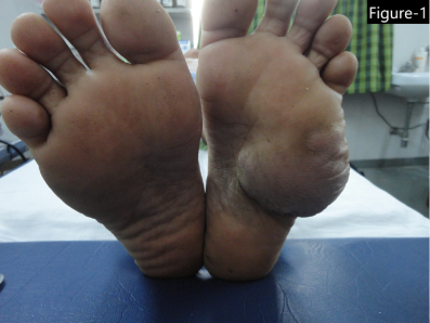
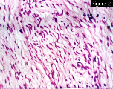
A 17 years old boy with normal IQ presented to the skin opd with a single, soft, elongated, non-tender swelling of size 6*4 cm over the sole of left foot Figure 1 ; with multiple caf´e-aulait: spots since childhood. Ocular examination showed lisch nodules. No other systemic abnormalities were found. Family history was negative. No history of consanguineous marriage. Routine investigations, USG of local part and CT scan were normal. The swelling was excised by a surgeon and histopathological examination was done, keeping the differentials as neurofibromatosis, lymphangioma, lipohemangioma. It showed interlacing bundles of elongated cells with wavy, darkly stained nuclei with cells were arranged in short fascicles at some places. The stroma showed wire-like strands of collagen (shredded carrots), areas of myxoid changes and vascular proliferation and adipose tissue intermingled with the tumour cells Figure 2: Based on these findings, it was diagnosed as PNF.
Case Report 2
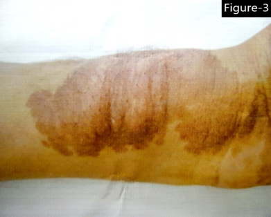
A 20 years old boy with normal IQ presented with a single, large, soft, elongated, non-tender swelling of size 10*5 cm over a pre-existing caf´e-aulait spot on ventral aspect of left forearm near the wrist Figure 3 : and multiple caf´e-aulait: macules all over the body. He had freckling over both palm and axilla. Ocular and other systemic examination was normal. Family history was negative, with no history of consanguineous marriage. Routine investigations, USG of local part and CT scan were normal. On basis of these clinical findings, the patient was diagnosed to have plexiform neurofibroma.
Discusion
Neurofibroma, a benign nerve sheath tumour in the peripheral nervous system. Out of its several forms, NF type 1 or von Recklinghausen's disease is the most common, presenting with many cutaneous manifestations. It arises from nonmyelinating -type schwann cells that exhibit inactivation of the NF-I gene, a tumour suppressor gene that codes for the protein neurofibromin3, leading to uncontrolled cell proliferation.4 Café-au-lait macules are well circumscribed, light brown macule or patch, 0.5 to 50 cm in size, present in 90% of cases. Freckles, the pathognomic feature of NF-1 appear later in 70% of the affected individuals (Crowe’s sign). 90% adults and 30% children younger than 6 years have pigmented iris hamartomas called Lisch nodules. Optic glioma, skeletal abnormalities including scoliosis, pseudarthrosis of tibia, mental retardation, pulmonic valvular stenosis, sexual precocity etc may occur. Cutaneous neurofibromas are soft tumours of varying size distributed over trunk and extremities. They can affect mucous membranes, visceral organs such as heart, lungs, brain and GIT. NF has four variants, the first type is characterized by discrete, blue-tinged cutaneous neurofibromas in epidermis and dermis. The second has subcutaneous neurofibroma as occurring deep in dermis, along the course of peripheral nerves, the third type consists of localized nodular PNF that interdigitate in normal tissues and the fourth type is characterized by diffuse plexiform neurofibromas (PNF). PNFs represent an uncommon variant (30%) of NF-1 but are one the most debilitating complications. NF-I is an autosomal dominant genetically inherited disorder, but only 50% patients of PNF have positive family history, the rest occur as spontaneous mutations. Uncontrolled proliferation of neural tissue in the subcutaneous fat, appears as bulging and deforming masses involving connective tissue and skin folds, giving it a ‘‘bags of worms’’ appearance. PNF can occur anywhere along a nerve and may appear on the abdomen,5 face,6 scalp, neck, chest, pelvis or spinal cord. The commonest site is along the trigeminal nerve distribution7 Many cases of facial PNF have been described. Singh P et. al have reported an unusual case of giant unilateral PNF of lumbosacral plexus extending into the thigh upto the level of knee joint. Aditi Mahalle et. al reported a rare case of solitary non syndromic oral PNF. It has a risk of malignancy, which ranges from 5 % to 28%.NF is clinically diagnosed on NIH (National institute of health consensus development conference) criteria, 1988 in which two or more cardinal signs are required - 6 or more brownish discolorations on the skin, more than 5mm in diameter in pre-adolescents and greater than 15 mm in adolescents and adults; 2 or more neurofibromas; freckling in the axilla or groin; optic glioma; 2 or more Lisch nodules or small masses oniris; bone lesions like sphenoid dysplasia; a first degree relative with neurofibromatosis 8. NF1-like syndrome, familial caf´e-aulait spots, segmental NF1 can be kept as differentials of PNF. Treatment of NF1 is mostly symptomatic. Large, disfiguring, or rapidly progressive and painful lesions can be excised. Several drugs are under trial for treating plexiform neurofibromas. Pirfenidone, a farnesyl transferase inhibitor used as an antifibrotic agent, downregulates the ras oncogene PEG, and is useful for this purpose.9 Gene therapy for the NF 1 gene represents the ultimate solution to prevent the wide range of manifestations caused by its mutation. 10
Conclusion
PNF is a hallmark lesion of NF type 1, its most common site face has been reported a lot of time in past. In our article we have presented 2 unusual cases of PNFs over the sole and forearm. PNF happens to be psychologically traumatic for most patients but in our case it let to more of a functional discomfort. Due to risk of malignant transformation in PNF, regular follow up is necessary.


