- Visibility 201 Views
- Downloads 6 Downloads
- DOI 10.18231/j.ijced.2020.063
-
CrossMark
- Citation
Wood’s lamp an antique but precious diagnostic tool: A descriptive observational study of fluorescence pattern with wood’s lamp in clinically diagnosed patients with pityriasis versicolor
- Author Details:
-
Arumugakani V
-
P K Kavirasan
-
Kannambal K
-
Poorana B
-
Abhirami C *
Introduction
Pityriasis versicolor (PV) is an innocuous, superficial non invasive fungal infective dermatosis in the worldwide. It is associated with alterations in pigmentation caused by normal inhabitant of skin flora named as Malassezia, a lipophilic dimorphic fungus. This fungus can cause disease when it converts to its pathogenic mycelia form under certain predisposed environmental, genetic, and immunological factors.[1]
Pityrialactone and pityriaanhydride are the flurochromes, primarily products of bisindolylcyclopentenetriones, unprecedented metabolites of Malassezia furfur. The first identified metabolite compound pityrialactone is a new fluorochrome bisindolyl compound which has both a bisindole and a lactone structure. Tryptophan is the main nitrogen molecular source of M. furfur , for the formation of fluorochromes. It was produced when M. furfur was incubated at 32 C for 14 days on a pigment inducing medium, and with the agar extract producing pale yellow compound eluting from the column which react with 22% acetonitrile and is found to exhibit a strong green-yellow fluorescence.[2]
Pityrialactone appears to be responsible for the green-yellow fluorescence of pityriasis versicolor lesions under Wood’s light. Its maximum absorption spectrum appears to be in the wavelength of 352, 292, 276 and 224 nm respectively. Pityriaanhydride which has not yet been described as a natural product but is a known intermediate in the total synthesis of bisindolylmaleimides. [2]
The individual color variation in fluorescence pattern of PV lesions, which may be explained interestingly by the variation does occurs in excitation spectrum and by the property of pityrialactone whether it was dissolved in water or acetonitrile. It fluorescence pale yellow in watery environment and blue in lipophilic milieu. As the skin contains both the solvents such as water and fats, the fluorescence varies. [2]
Woods light physics
The Wood's light (“black light”), first described in 1903, in early 20th century, by an American Baltimore physicist Robert Williams Wood (1868–1955). Though several types of Wood's lamps are available, the most inexpensive ones being the hand models with batteries and small fluorescent tubes, with a high-intensity 100-watt mercury vapor bulb and power supply. It work on the basic principle of fluorescence. [3] The first reported use of this lamp in dermatology occurred in 1925, being recommended for the detection of fungal infection of the hair done by Margarot and Deveze. [4]
When subjecting the skin to wood’s lamp examination, a high-pressure mercury arc generate long-wave UV radiation (UVR), the arc is fitted with a compounded filter made of barium silicate with 9% nickel oxide, the so-called ‘Wood’s filter which is opaque to all generated light except for a band between 320 and 400 nm with a peak at 365 nm. This generated shorter wavelengths is absorbed in the diseased skin and radiation of longer wavelengths (400-700nm) usually visible light, is emitted from the skin and finally fluorescence of tissue occurs.The output of Wood’s lamp is generally low. [5] As tissue autofluorescence appears to derive mainly from constituents of elastin (fluorophore unknown), collagen (pyridinoline crosslinks), aromatic amino acids (predominantly tryptophan and its oxidative products), nicotinamide adenine dinucleotide (NAD), and perhaps precursors or products of melanin causing bluish fluorescence. [6]
Materials and Methods
This study was an observational descriptive study, conducted in 100 clinically suspected cases of Pityriasis Versicolor who attended the department of Dermatology, Venereology and Leprosy at Rajah Muthaiah Medical College, over a period of 1 year (October 2019 to October 2020). Ethical clearance was sought from Institutional Ethical Committee. Written informed consent was obtained, the relevant epidemiological data was taken in relation to age, sex, duration of illness and recurrent infections. Clinical examination of lesion included number, site, types of lesions and scaling. All the PV patients were evaluated under Wood’s lamp examination with the fulfilment of a few requirements. The first and foremost obvious requirement is a completely darkened room for the investigation. Second, the user should be aware that the human eye needs some time to adjust to the low light environment in which the investigation takes place. Wood’s lamp should be on 20 sec to one minute and kept at the distance of four inches from examination site.
Exclusion criteria
Those who have applied topical oil, turmeric, medicated onintments.
Those who are on treatment with specific anti fungal therapy less than 4 weeks duration.
Observations and Results
The study group was comprised of 100 clinically diagnosed cases of Pityriasis Versicolor. Among the 100 patients, 53 were males and the remaining 47 were females with a male to female ratio of 1.12:1.
|
Age in years |
Sex |
No of cases |
Percentage |
|
|
male |
Female |
|||
|
Less than 1-10 |
6 |
12 |
18 |
18% |
|
11-20 |
19 |
11 |
30 |
30% |
|
21-30 |
17 |
11 |
28 |
28% |
|
31-40 |
3 |
6 |
9 |
9% |
|
41-50 |
4 |
5 |
9 |
9% |
|
51-60 |
4 |
2 |
6 |
6% |
|
|
53 |
47 |
100 |
100% |
In our study majority of the age group with PV were observed 11-20 were 30 (30%), followed by 21-30 were 28 (28%), less than one year and 10 years were 18 (18%), 31-40 years were 9(9%), 41-50 years were 9(9%), 51-60 years were 6(6%).
|
site of skin lesions |
Number of sites |
|
Upper chest |
45 |
|
Back |
39 |
|
Face |
38 |
|
Neck |
17 |
|
upper extremities |
11 |
|
Flexural |
3 |
|
lower extremities |
3 |
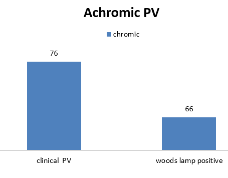
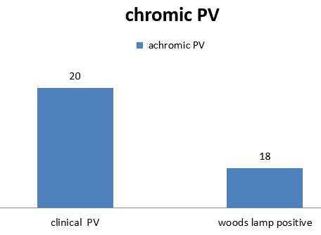
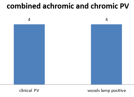
Patients presented with duration of 1–6 months 64(64%), 6 months to 1 year were 24(24%), one year to one and half years were 12(12%). Patients presented with itching were 15(15%), remaining were not have itching. Patients presented with scaling were 87(87%), 13 cases were not with scales. There was a history of family members having similar complaints in 13(13%), and in 87(87%) there was no family history.
|
Type of skin lesions |
Total no of cases |
percentage |
New cases |
percentage |
Recurrence |
percentage |
|
Achromic |
76 |
76% |
47 |
47% |
29 |
29% |
|
Chromic |
20 |
20% |
11 |
11% |
9 |
9% |
|
Both |
4 |
4% |
2 |
2% |
2 |
2% |
|
Total |
100 |
100% |
60 |
60% |
40 |
40% |



Discussion
In our study, the lesions were most commonly seen in the age group of 11-20 (30%) years, as has been reported by other authors. Ghosh et al [7] found the incidence to be 32.37(32.37%) in young adults in the age group 12-21 years, and Rao et al [8] found in their study that 30% of the patients belonged to the age group of 21-30 years. The disease may occur at any age, but it is more common during adolescence and in young adults due to an increase in the sebaceous activity and hormonal changes. The present study showed a pre-dominant involvement of males (53%) as compared to females(47%) with the ratio of 1.12:1 correlated with Ghosh et al.[7] The higher incidence of Pityriasis versicolor in males may be due to their outdoor activities.
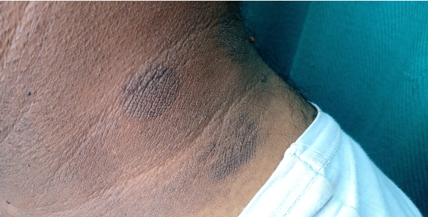
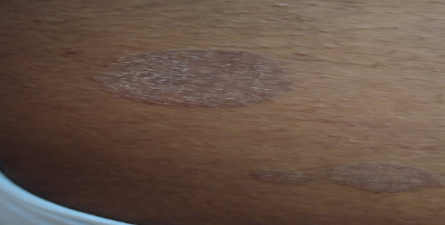
The type of lesions were more achromic (76%) than chromic(20%), which was similar to the study of Rao et al. [8] The distribution of the skin lesions commonly occurred over the chest (45%), and the upper back (39%). These observations were not in conformity with the findings of Rao et al [8] who found that the lesions were seen commonly over the back (70%) and the chest (58.30%). The distribution of the lesions generally parallels the density of the sebaceous gland distribution, with a greater occurrence on the chest and the back.
In our study yellow fluorescence observed in patients who were clinically suspected case of Pityriasis Versicolor which was concordance with previous literature yellow fluorescence in PV reported by Robert Williams [9] and many authors. In our study 86(86%) of patients showed PV yellow fluorescence under Wood’s lamp examination, as has been reported by Remya et al [10] who compared Wood‟s lamp examination with KOH wet mount in patients with PV. They concluded that Wood‟s lamp positive in 86.2% of the PV lesions studied.
Wood’s lamp examination for showing yellow-orange fluorescence was found to be positive in 70% of the cases reported by Deshpande SA et al. [11]
There was no fluorescence detected in 10(10%) cases of infants less than 1 year and children upto 3 years who were included in the study. Fahimeh et al [12] reported that Wood’s lamp observation in infantile pityriasis versicolor some lesions of hypopigmented areas showed yellowish fluorescence .
In these group face was the commonest site which was similar to the previous study conducted by Terrangni et al. [13] Hypochromic was the commonest clinical presentation in paediatric Pityriasis Versicolor which was similar to the study reported by Bouassida et al. [14]
Wood’s lamp is equally effective in detecting new and recurrent cases of PV. Intensity or brightness of yellow fluorescence detected highly in chromic PV skin lesions than achromic PV under Wood’s lamp may be the reason due to increased number of spores and hyphae, abnormally large melanososmes and melanin granules, and induction of melanogenesis. [15] Wood’s lamp negativity 14 cases (14%) may be explained by different species of malassezia [15] and the variation in the production of fluorochromes.
Conclusion
Though the mycological study in Pityriasis versicolor by doing Potassium hydroxide mount or by other conventional methods is simple, it is impossible to do in the situations like non scaly and atypical presentation without suspicious of Pityriasis versicolor. We can easily avoid misdiagnosis by usage of Wood’s lamp in daily out patient department.
Limitations
Chance of unwarranted diagnosis by wrong interpretation of scales which fluoresce normally as bluish white. Total removal of scales in infective conditions abolishes the fluorescence pattern and may give false negative results. So we need a experience to interpret the results.
Source of Funding
No financial support was received for the work within this manuscript.
Conflict of Interest
The authors declare they have no conflict of interest.
References
- S. Renati, A. Cukras, M. Bigby. Pityriasis versicolor. BMJ 2015. [Google Scholar] [Crossref]
- P Mayser, H Stapelkamp, H-J Krämer, M Podobinska, W Wallbott, B Irlinger. Pityrialactone- a new fluorochrome from the tryptophan metabolism of M. alassezia furfur. Antonie Van Leeuwenhoek 2003. [Google Scholar] [Crossref]
- E Ruocco, A Baroni, G Donnarumma, V Ruocco. Diagnostic procedures in dermatology. Clin Dermatol 2011. [Google Scholar] [Crossref]
- J Margarot, P Deveze. Aspect de quelques dermatoses en lumie`re ultraparaviolette. Note pre´liminaire. Bull Soc Sci Med Biol Montpellier 1925. [Google Scholar]
- R M Caplan. Medical Uses of the Wood's Lamp. J Am Acad Dermatol 1967. [Google Scholar]
- P Asawanonda, C R Taylor. Wood's light in dermatology. Int J Dermatol 1999. [Google Scholar] [Crossref]
- S K Ghosh, S K Dey, I Saha, J N Barbhuiya, A Ghosh, A K Roy. Pityriasis versicolor: A clinicomycological and epidemiological study from a tertiary care hospital. Indian J Dermatol 2008. [Google Scholar] [Crossref]
- G S Rao, M Kuruvilla, P Kumar, V Vinod. Clinico Epidemiological studies on tinea versicolor. Indian J Dermatol Venereol Leprol 2002. [Google Scholar]
- S Sharma, A. Robert Williams Sharma, Wood. Robert Williams Wood: pioneer of invisible light. Photodermatol Photoimmunol Photomed 2016. [Google Scholar]
- V S Remya, B Arun. Diagnostic Efficacy of Wood’s Lamp Examination Compared with Koh Wet Mount for Diagnosis of Pityriasis Versicolor Cases. Int J Hea Sci Research 2019. [Google Scholar]
- S A Deshpande, D Sunita, S Amladi, J Shastri. The role of mycological investigations in the diagnosis of Pityriasis Versicolor and Seborrheic dermatitis. Ind J Bas App Med Research 2014. [Google Scholar]
- F Abdollahimajd. Infantile hypopigmented pityriasis versicolor: two uncommon cases. Türk Pediatri Arşivi 2019. [Google Scholar] [Crossref]
- L. Terragni, A. Lasagni, A. Oriani, C. Gelmetti. Pityriasis Versicolor in the Pediatric Age. Mycoses 1991. [Google Scholar] [Crossref]
- S Bouassida, S Boudaya, R Ghorbel, T J Meziou, S Marrekchi, H Turki, A Zahaf. Pityriasis versicolor de l'enfant: étude rétrospective de 164 cas Pityriasis versicolor in children: a retrospective study of 164 cases. Ann Dermatol Venereol 1998. [Google Scholar]
- D M Thappa, D Gupta. The enigma of color in tinea versicolor. Pigment Int 2014. [Google Scholar] [Crossref]
