- Visibility 88 Views
- Downloads 8 Downloads
- DOI 10.18231/j.ijced.2024.075
-
CrossMark
- Citation
Comparative evaluation of intralesional platelet rich plasma versus tranexamic acid (50mg/ml) in the treatment of facial melasma
Introduction
Melasma, also referred to as chloasma, is a common hyperpigmentary disorder characterized by brown patches on the face, typically affecting middle-aged women. [1] Several contributing factors have been identified, including genetic predisposition, prolonged sun exposure, pregnancy, contraceptive use, medications, and hormonal therapy. [2] Furthermore, the development of melasma may be attributed to dysfunction in melanogenesis, involving the transfer of melanosomes to keratinocytes through the tyrosinase enzyme, which regulates melanin production. [3] Various treatment options, such as topical depigmenting agents, chemical peels and laser therapies [4] have been explored in various research studies, yielding inconsistent and often unsatisfactory results. Platelet-rich plasma (PRP) refers to a small amount of plasma derived from the patient's own blood, which contains a high concentration of platelets. The presence of platelet derived growth factors promote angiogenesis and collagen synthesis, leading to an increase in skin volume and improvement in melasma. [5] Additionally, the release of transforming growth factor beta (TGF-β1) from platelet α-granules has been found to significantly inhibit melanin synthesis by delayed extracellular signal-regulated kinase activation. [6] Tranexamic acid (TXA), a traditional hemostatic drug has hypopigmentory effect on melasma lesions and also prevents ultraviolet induced pigmentation.[7] The intracellular release of arachidonic acid as precursor of prostanoid and the level of alpha-melanocyte stimulating hormone increase as the result of plasmin activity. These two substances can activate melanin synthesis. Therefore, the anti-plasmin activity of tranexamic acid is thought as the main mechanism of hypopigmentary effect of this agent.[8] Studies comparing PRP and TXA in a higher concentrations (50mg/ml) are limited. Hence this study has been undertaken to evaluate the therapeutic efficacy and safety of PRP and TXA in a higher concentration (50mg/ml).
Materials and Methods
Study population and sample size
This study was performed after obtaining approval from the Institutional Ethical Committee (approval number: SGRD/IEC/22-138). A total of 66 clinically diagnosed cases of facial melasma from July 2023 to March 2024, visiting to the outpatient department of Dermatology, Venereology and Leprosy at a tertiary care centre in North India, were enrolled in the study after taking informed written consent. All patients were randomly divided into 2 groups (A& B), out of which 2 patients from group A left the study in between and excluded from the study. In group A, 31 patients were treated with intralesional PRP and in group B, 33 patients were treated with intralesional TXA (50mg/ml).
Exclusion criteria
The study did not include patients who had a history of bleeding disorders and thromboembolic events, abnormal coagulation profile, simultaneous use of anticoagulants and photosensitizing medications, previous history of taking OCP’s or hormonal therapies within last 1 year, known allergy to components of sunscreen or topical anaesthetic used in this trial, any active infection at the injection site, history of chemotherapy and radiotherapy, tendency to form hypertrophic scar/keloid, pregnancy and lactation.
Requirement and procedure
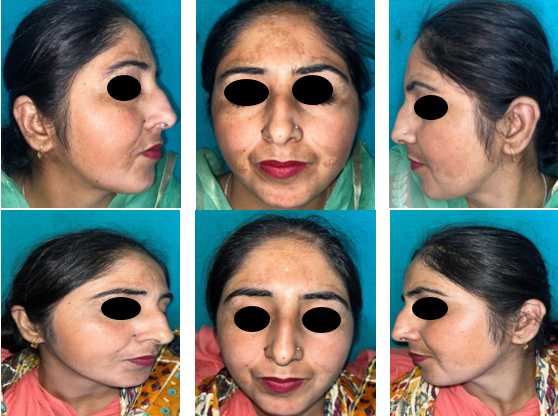
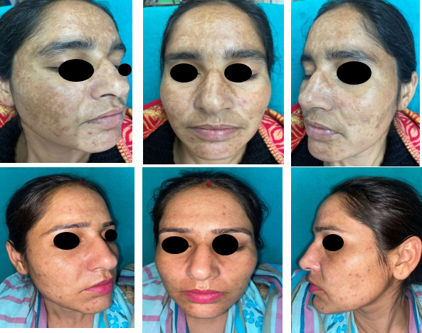
All the patients were explained about the procedure in detail. Thorough history was taken and a comprehensive dermatological evaluation was conducted. Fitzpatrick skin type and clinical pattern (centrofacial, malar or mandibular) of melasma were noted in all patients. Morphological type (epidermal, dermal or mixed) of melasma was also assessed by using ILLUCO IDS 1100 dermatoscope. The lesions were captured digitally before and after the completion of the treatment using the 12 MP camera of an Apple iPhone 11. All enrolled patients were advised to get complete blood count, bleeding time, clotting time, viral markers and thyroid profile done at the beginning. Patients were advised to wash their face thoroughly with water and mild cleanser. Topical anaesthetic cream (containing 2.5% prilocaine & 2.5% lidocaine) was applied on face for 45 minutes under occlusion before procedure and then wiped off to obtain completely dry skin.
PRP preparation
The PRP was prepared using the double-spin method. A total of 20 mL of the patient's blood was collected in a sterile vacutainer tube containing anticoagulant acid citrate dextrose-A. Initially, a slow spin was carried out at 1000 revolutions per minute for 10 minutes at room temperature. Following the first spin, the plasma was carefully aspirated and transferred to a second set of vacutainers. A second spin (heavy spin) was then conducted at room temperature at a rate of 2000 RPM for 5 minutes. The upper two-thirds of this double spinned plasma was collected as platelet-poor plasma, while the bottom one-third was utilized as PRP.
TXA preparation
A 0.5ml injection of TXA (500mg/5ml) was drawn in an insulin syringe and then diluted with 0.5 ml of normal saline to achieve a concentration of 50mg/ml TXA. 0.05 ml of the solution was injected intralesionally through a 30 gauze needle insulin syringe at 1cm intervals on the facial melasma lesion. The maximum permissible dose in a single session was 250 mg.
In group A, patients were treated with 0.1 ml intralesional PRP injection while in group B, 0.05ml of intralesional TXA was given through a 30 gauge insulin syringe in each cm2 of melasma. This procedure was repeated every 4 weekly till lesions resolved or a maximum of 6 sessions followed by monthly follow up for 3 months after last treatment session. Following the procedure, all patients were directed to apply broad-spectrum sunscreen with SPF 30. Serial photography and mean mMASI score were utilized to evaluate the clinical response at baseline, during each session, and at every follow-up. Throughout the study and at each subsequent follow-up, patients were closely monitored for any adverse events and managed accordingly. The efficacy of the treatment was evaluated by calculating the percentage reduction in mean mMASI during each session and at every subsequent follow up. An independent observer, who was not involved in the study, compared the photographs taken before and after the treatment. Furthermore, the patient satisfaction score was assessed using a five-point Likert scale.
Statistical analysis of data
The data was entered into MS Excel 2013. Collected data was then analyzed using Statistical Package for Social Sciences (SPSS) version 23.0. The comparison between groups was made using the Chi-square test. For comparing two independent groups with parametric quantitative variables, the independent samples t-test was used, and the Mann-Whitney U test was used for non-parametric data. A p-value below 0.05 was considered statistically significant and p-value below 0.001 was considered statistically highly significant.
Results
There were total 55 females (85.94%) patients and 9 males (14.06%). The age range in our study was from 23 to 47 years with mean age of 34.67±6.71 years, but most of the study participants were in the age group of 31 to 40 years (42.18%). All the patients included were having Fitzpatrick skin type 3 or 4. The mean duration of the disease among enrolled cases was 7.85±4.07 years with range between 2 years and 12 years. Out of 64 patients a positive family history of melasma was present in 18 cases (28.125%) and 56 patients (87.5%) had a history of chronic and persistent sun exposure. 41 cases (64.06%) had centrofacial pattern, 12 cases (18.75%) had malar pattern and only 11 cases (17.19%) presented with mandibular pattern of distribution of melasma. Among the total 64 participants, epidermal, dermal and mixed pattern was seen in 13, 16 and 35 patients respectively on dermatoscopic examination. ([Table 1]).
In group A mean mMASI score at baseline was 6.794±3.639 and it was reduced to 3.710±2.595 after last follow up and the percentage reduction in mean mMASI was 46.596% from baseline. In group B mean mMASI score decreased from a baseline value of 5.452±3.380 to 2.224±1.797 with a 58.131% percentage reduction in mean mMASI. There was a statistically significant decline in the mMASI score between both the groups (p=0.006) at last follow up and percentage reduction in mean mMASI score was higher in group B as compared to group A ([Table 2]).
There were 2 males and 29 females patients in group A. No significant reduction in mean mMASI score was observed in male patients from baseline to last follow up in group A (PRP). From baseline to last follow up, a highly significant (p=0.0001) reduction in mean mMASI score was found in female patients treated with intralesional PRP. There were 7 male and 26 female patients in group B. Significant reduction (p=0.0037) in mean mMASI score was observed in male patients from baseline to last follow up after treatment with intralesional TXA whereas this reduction was highly significant (p=0.0001) in female patients when treated with intralesional TXA in similar time line.([Table 3])


|
Demographic |
No. of cases |
%age |
|
Gender |
|
|
|
• Male |
9 |
14.06 |
|
• Female |
55 |
85.94 |
|
Age group |
|
|
|
• 20-30 |
23 |
35.93 |
|
• 31-40 |
27 |
42.18 |
|
• 41-50 |
14 |
21.87 |
|
Mean age |
34.67±6.71 |
|
|
Fitzpatrick skin type |
|
|
|
• I |
0 |
0.00 |
|
• II |
0 |
0.00 |
|
• III |
45 |
70.31 |
|
• IV |
19 |
29.68 |
|
• V |
0 |
0.00 |
|
• VI |
0 |
0.00 |
|
Duration of disease |
|
|
|
• <5 years |
22 |
34.38 |
|
• 5-10 years |
20 |
31.25 |
|
• >10 years |
22 |
34.38 |
|
Mean duration of disease |
7.85±4.07 |
|
|
Family history |
|
|
|
• Absent |
46 |
71.875 |
|
• Present |
18 |
28.125 |
|
Sun exposure |
|
|
|
• Absent |
8 |
12.500 |
|
• Present |
56 |
87.500 |
|
Clinical pattern of melasma |
|
|
|
• Centrofacial |
41 |
64.06 |
|
• Malar |
12 |
18.75 |
|
• Mandibular |
11 |
17.19 |
|
Morphological pattern of melasma |
|
|
|
• Epidermal |
13 |
20.313 |
|
• Dermal |
16 |
25.000 |
|
• Mixed |
35 |
54.688 |
|
Time interval |
Mean mMASI score |
||||
|
Group A (n=31) PRP |
Group B (n=33) TXA |
p-value |
|||
|
Mean |
SD |
Mean |
SD |
||
|
Before treatment |
6.794 |
3.639 |
5.452 |
3.380 |
0.082 |
|
After treatment |
3.710 |
2.595 |
2.224 |
1.797 |
0.006 |
|
Mean %age reduction |
46.596 |
22.611 |
58.131 |
21.672 |
0.037 |
|
Time interval |
Group A |
Group B |
||||||
|
Male (n=2) |
Female (n=29) |
Male (n=7) |
Female (n=26) |
|||||
|
Mean mMASI |
SD |
Mean mMASI |
SD |
Mean mMASI |
SD |
Mean mMASI |
SD |
|
|
Baseline |
7.90 |
0.98 |
6.71 |
3.74 |
5.92 |
2.83 |
5.32 |
3.55 |
|
At last follow up |
5.55 |
4.03 |
3.58 |
2.52 |
1.88 |
0.92 |
2.31 |
1.97 |
|
p-value |
0.507 |
0.0001 |
0.0037 |
0.0001 |
|
Time interval |
Group A |
Group B |
||||||||||
|
Epidermal |
Dermal |
Mixed |
Epidermal |
Dermal |
Mixed |
|||||||
|
Mean mMASI |
SD |
Mean mMASI |
SD |
Mean mMASI |
SD |
Mean mMASI |
SD |
Mean mMASI |
SD |
Mean mMASI |
SD |
|
|
Baseline |
5.56 |
2.67 |
6.78 |
3.77 |
7.20 |
3.94 |
3.60 |
2.54 |
6.49 |
4.52 |
5.67 |
2.84 |
|
At last follow up |
3.47 |
2.34 |
4.50 |
3.89 |
3.48 |
2.15 |
1.17 |
1.16 |
2.48 |
1.93 |
2.52 |
1.87 |
|
p-value |
0.17 |
0.28 |
0.001 |
0.04 |
0.02 |
0.0006 |
|
Grade of clinical improvement |
Group A |
Group B |
||
|
No. |
%age |
No. |
%age |
|
|
Very good (>75% reduction in mean mMASI score) |
3 |
9.68 |
10 |
30.30 |
|
Good (51-75% reduction in mean mMASI score) |
10 |
32.26 |
14 |
42.42 |
|
Moderate (25-50% reduction in mean mMASI score) |
10 |
32.26 |
3 |
9.09 |
|
Mild (<25% reduction in mean mMASI score) |
6 |
19.35 |
5 |
15.15 |
|
No (no reduction in mean mMASI score) |
2 |
6.45 |
1 |
3.03 |
|
Total |
31 |
100.00 |
33 |
100.00 |
|
P-Value |
X2: 8.575; df:4; p=0.073 |
|
Score on Likert scale |
Group A |
Group B |
||
|
No. |
%age |
No. |
%age |
|
|
1 (Not at all satisfied) |
2 |
6.45 |
1 |
3.03 |
|
2 (Not really satisfied) |
6 |
19.35 |
2 |
6.06 |
|
3 (Undecided) |
10 |
32.26 |
3 |
9.09 |
|
4 (Somewhat satisfied) |
10 |
32.26 |
16 |
48.48 |
|
5 (Very much satisfied) |
3 |
9.68 |
11 |
33.33 |
|
Total |
31 |
100.00 |
33 |
100.00 |
|
P-Value |
X2: 12.008; df:4; p=0.017 |
In group A, 6 patients had epidermal pattern, 7 patients had dermal pattern and 18 patients had mixed pattern of melasma. No significant reduction in mean mMASI score was noticed in epidermal as well as dermal pattern of melasma after treatment with intralesional PRP from baseline but significant reduction (p=0.001) in mean mMASI score from baseline after treatment with intralesional PRP was found in mixed pattern of melasma. There were 7 patients with epidermal pattern, while 9 and 17 cases had dermal and mixed patterns respectively in group B. Significant reduction in mean mMASI score from baseline to last follow up was noted in epidermal (p=0.04) as well as dermal (p=0.02) patterns while this reduction was highly significant in mixed pattern (p=0.0006) of melasma from baseline to last follow up. ([Table 4])
In the PRP group, 64.52% patients showed moderate (25-50% reduction in mMASI score) to good (51-75% reduction in mMASI score) response whereas in the TXA group, 72.72% patients showed good to very good (>75% reduction in mMASI score) response as per grade of clinical improvement ([Table 5]).
In group A, only 41.94% patients were satisfied with the treatment while in group B, majority of the patients (81.81%) were satisfied with their recovery after treatment as per patient satisfaction score on 5 point Likert scale. ([Table 6])
Discussion
Melasma is a common distressing condition for patients as well as for dermatologists due to its recalcitrant nature. It has a negative impact on the patient’s quality of life. As a result, melasma continues to be a difficult condition that requires a novel and effective treatment approach.
In the present research, the female to male ratio was 6.1:1. Similarly the researches conducted by Achar A and Rathi SK,[9] Serra et al,[10] and Jin et al,[11] also stated that females outnumbered males, thus reiterating that melasma is more frequently seen in females. The mean age of participants in the current study was 34.67±6.71 years. Sarkar et al,[12] also reported a mean age of 30.7 years in their study. This could be because melasma is more common in reproductive age group due to hormonal imbalance. All patients in the present study had fitzpatrick skin types III and IV. Farag MM et al,[13] also observed that all individuals involved in their study exhibited Fitzpatrick type III and IV. The current study revealed that the average duration of the disease among the participants was 7.85±4.07 years, with a range of 2 to 12 years. Kumar S et al[14] in 2014, reported an average duration of melasma of 1.79 years, indicating a lower duration compared to other studies According to current study’s observation, centrofacial pattern (64.06%) was the most common clinical pattern among the cases while mandibular pattern was least common (17.18%). Similar pattern was found in a study by Fariyal Hoque et al.[15] which stated that 76% of all melasma variants had centrofacial pattern as the most common clinical pattern. Among the morphological pattern, mixed pattern of melasma was observed as the most common (54.68%) in the present study followed by dermal pattern (25%) and epidermal pattern (20.31%). It could be due to the fact that epidermal melasma responds well to local treatment, whereas dermal and mixed types are less or non-responsive.
In group A mean mMASI score reduced from 6.794+_3.639 at baseline to 3.710=-2.595 at the last follow up visit and the percentage reduction in mean mMASI was 46.596% from baseline. In group B mean mMASI score decreased from a baseline value of 5.452±3.380 to 2.224±1.797 with a 58.131% percentage reduction in mean mMASI. There was a statistically significant decline in the mean mMASI score between both the groups after treatment (p=0.006) and the percentage reduction in mean mMASI score was higher in group B as compared to group A (p=0.037). Lal M et al,[16] in their study on intralesional TXA (4mg/ml) versus PRP in melasma observed a significant reduction in mean mMASI score in both the groups but the comparison of overall improvement in both the groups was not significant. Similarly in a split face study done by Abd Elraouf IG et al[17] statistically significant reduction in mean mMASI score was seen with both PRP and TXA, but a higher percentage reduction in mean mMASI score was observed with PRP. Mumtaz et al,[18] also observed more favourable results with intradermal PRP for treatment of melasma. The better results with TXA in the present study, could be attributed to the greater number of visits and a higher TXA concentration (50mg/ml).
The current study observed statistically highly significant reduction (p=0.0001) in mean mMASI score in female patients with PRP as well as TXA. This could be due to the fact that women are cosmetically more concerned, start early treatment, maintain regular follow up, adhere properly to treatment and have a lesser tendency for sun exposure. In the study, a significant reduction (p=0.0037) in mean mMASI score from baseline to last follow up was also noted in male patients after treatment with TXA. Similar results with TXA were also found in a study by Verma YR et al,[19] This similarity could be attributed to higher number of treatment sessions with TXA which was used in both the studies. Most of the other available studies on TXA did not compare therapeutic outcome between male and female patients in melasma.
After a comprehensive analysis of data at last follow up, it was found that efficacy of PRP was statistically significant in mixed melasma (p=0.001). Similar results were obtained in a study by Hofny et al,[20] in which three monthly sessions of PRP were administered and a significant reduction in the mean mMASI score (p<0.05) was observed in mixed type of melasma. In the present study, reduction in mean mMASI score from baseline to last follow up was statistically highly significant with TXA in mixed pattern (p=0.0006) and significant in epidermal as well as dermal melasma (p<0.05). Our results are concordant with the study by Verma YR et al[19] in epidermal and mixed patterns but in contrast when compared to dermal melasma. The better response, even in dermal melasma, in the present study could probably be attributed to the higher concentration of TXA(50mg/ml) used as compared to 25mg/ml TXA used in the study by Verma Y R et al.[19] At last follow up, the authors found PRP to be most effective in centrofacial melasma while the TXA group showed best results in mandibular distribution of melasma and p value was 0.0034 (statistically highly significant reduction in mean mMASI score). Significant reduction (p<0.05) in mean mMASI score was also noted in centrofacial and malar distribution of melasma with TXA. It is important to note that none of the other available research studies emphasized on treatment response variations among different morphological patterns of melasma. In the current study, moderate to good response was noticed in 64.52% patients with PRP whereas good to very good response was noticed in 72.72% patients of TXA group. However, these results were not statistically significant (P value was 0.073). Similar results were seen in a split face comparative study between intralesional TXA (4mg/ml) and PRP by Lal M et al, [16] where intralesional TXA demonstrated superior improvement as compared to PRP in treating melasma but results were not significant (p<0.05). The authors noticed 41.94 % satisfied patients after treatment in group A compared to 81.81% in group B (p=0.017). This difference was statistically insignificant in study by Abd Elraouf IG et al. [17] The authors also noted that all patients experienced common side effects including pain and erythema at site of injection during each treatment session, which eventually disappeared within a few hours. No serious local or systemic adverse effects were observed.
Conclusions
Melasma is a persistent and recurrent disorder of hyperpigmentation. While many different therapeutic modalities have been explored in the past, none of them have produced satisfying outcomes. As a result, melasma continues to be a difficult condition in need of novel and efficient treatment approaches. Our results suggest that intralesional PRP and TXA (50mg/ml) were both linked to a statistically significant decline in the mMASI score; however, the intralesional TXA group showed a greater percentage reduction in mMASI score and an earlier significant reduction in mMASI than the PRP group. TXA in higher concentration was effective in all clinical and morphological patterns of melasma. Patient satisfaction following treatment was also higher in group B compared to group A, and there was no statistically significant difference in the side effects between the two groups. We suggest monthly therapy with intralesional TXA at a higher dosage of 50 mg/ml as an efficient and time-saving treatment modality over intralesional PRP.
Source of Funding
None.
Conflict of Interest
None.
References
- S Rajanala, Mbc Maymone, NA Vashi. Melasma pathogenesis: a review of the latest research, pathological findings, and investigational therapies. Dermatol Online J 2019. [Google Scholar]
- BW Lee, RA Schwartz, CK Janniger. Melasma. G Ital Dermatol Venereol 2017. [Google Scholar]
- R Sarkar, P Ailawadi, S Garg. Melasma in men: a review of clinical, etiological, and management issues. J Clin Aesthet Dermatol 2018. [Google Scholar]
- L Zhao, M Hu, Q Xiao, R Zhou, Y Li, L Xiong. Efficacy and Safety of Platelet-Rich Plasma in Melasma: A Systematic Review and Meta-Analysis. Dermatol Ther (Heidelb) 2021. [Google Scholar]
- R Sarkar, A Bansal, P Ailawadi. Future therapies in melasma: What lies ahead?. Indian J Dermatol Venereol Leprol 2020. [Google Scholar]
- M Pistor. What is mesotherapy?. Chir Dent Fr 1976. [Google Scholar]
- HJ Kim, SH Moon, SH Cho, JD Lee, HS Kim. Efficacy and safety of tranexamic acid in melasma: a meta-analysis and systematic review. Acta Derm Venereol 2017. [Google Scholar]
- JY Chung, JH Lee, JH Lee. Topical tranexamic acid as an adjuvant treatment in melasma: side-by- side comparison clinical study. J Dermatolog Treat 2015. [Google Scholar]
- A Achar, SK Rathi. Melasma: a clinico-epidemiological study of 312 cases. Indian J Dermatol 2011. [Google Scholar]
- M Serra, K Bohnert, M Narda, C Granger, N Sadick. Brightening and improvement of facial skin quality in healthy female subjects with moderate hyperpigmentation or dark spots and moderate facial aging. J Drugs Dermatol 2018. [Google Scholar]
- Y Jin, W Jiang, Y Yao, H Huang, J Huang. Clinical efficacy of laser combined with menstrual regulation in the treatment of female melasma: a retrospective study. Lasers Med Sci 2019. [Google Scholar]
- R Sarkar, P Puri, R K Jain, A Singh, A Desai. Melasma in men: a clinical, aetiological and histological study. J Eur Acad Dermatol Venereol 2010. [Google Scholar]
- M M Farag, F F Mostafa, K M Gharib. Therapeutic effect of dermapen with PRP versus dermapen with tranexamic acid in melasma cases. Zagazig Univ Med J 2020. [Google Scholar] [Crossref]
- S Kumar, BB Mahajan, N Kamra. Melasma in North Indians: A clinical, epidemiological, and etiological study. Pigment Int 2014. [Google Scholar]
- F Hoque, J Mcgrath, SE Shaude. Melasma (Chloasma): Pathogenesis and Treatment. J Biotechnol Biomed 2022. [Google Scholar]
- M Lal, M Nijhawan, D Yadav, M Rijhwani, S Awasthi. A study of intralesional tranexamic acid versus platelet rich plasma in melasma - a split face comparative study. JOJ Dermatol and Copsmet 2021. [Google Scholar] [Crossref]
- IGA Elraouf, ZM Obaid, I Fouda. Intradermal injection of tranexamic acid versus platelet-rich plasma in the treatment of melasma: a split-face comparative study. Arch Dermatol Res 2023. [Google Scholar]
- M Mumtaz, TH Chandio, MK Shahzad, N Hanif, S Anwar, S Rafique. Comparing the Efficacy of Patelet-rich Plasma (PRP) versus Tranexamic Acid (4mg/mL) as Intradermal Treatments of Melasma. J Coll Physicians Surg Pak 2021. [Google Scholar]
- YR Verma, KS Mehta, PS Chauhan, VK Mahajan, M Chandel, HK Sharma. Study of the therapeutic efficacy and safety of Intralesional Tranexamic Acid (25 mg/ml) for the treatment of melasma in male patients: A single centered “beforeafter” observational study. Int J Res Dermatol 2021. [Google Scholar]
- ER Hofny, AA Abdel-Motaleb, A Ghazally, AM Ahmed, MR Hussein. Platelet-rich plasma is a useful therapeutic option in melasma. J Dermatolog Treat 2019. [Google Scholar]
