- Visibility 52 Views
- Downloads 6 Downloads
- DOI 10.18231/j.ijced.2024.079
-
CrossMark
- Citation
Dermatological manifestations and associated factors in patients with Graves' disease in Dakar
- Author Details:
-
Assane Diop *
-
Saer Diadie
-
Mame Tene Ndiaye
-
Mame Fatou Faye
-
Ismail Tounsi
-
Aby Seck
-
Demba Diédhiou
-
Boubacar Ahi Diatta
-
Maodo Ndiaye
-
Abdoulaye Lèye
-
Fatimata Ly
-
Suzanne Oumou Niang
Introduction
Dermatological manifestations of thyroid dysfunctions are often observed in Graves' disease, which remains the most common and expressive cause of thyroid hyperfunctioning.[1] These manifestations generally occur after the development of thyroid disease but can also be the first sign.[2] As such, they have diagnostic, therapeutic, and prognostic value.
Their mechanisms are complex and demonstrate the strong influence of thyroid hormones on the skin. A better understanding of their pathophysiology can allow for earlier diagnosis and improved management.[3] In Asia, a Malaysian study showed that one-fifth of hyperthyroid patients with Graves' disease presented dermatological manifestations.[4] In the Maghreb, a Tunisian study reported the presence of at least one dermatological sign in a series of hyperthyroid patients.[5]In sub-Saharan Africa, very few studies have been conducted on dermatological manifestations in Graves' disease.
Therefore, the objective of our work was to describe the epidemiological and clinical profile of dermatological manifestations in patients with Graves' disease and the associated factors.
Materials and Methods
This cross-sectional study involved prospective data collection conducted between March 1st and August 31st, 2021. The research was conducted within the endocrinology units of Clinique Médicale II at Centre Hospitalier Abass Ndao and the Internal Medicine/Endocrinology-Diabetology-Nutrition Department at Centre Hospitalier National de Pikine (CHNP).
Our study encompassed all individuals aged 18 years or older, irrespective of gender, undergoing follow-up for dysthyroidism. Specifically focusing on Graves' disease, we included patients aged 18 years or older, regardless of the presence of cutaneous, mucosal, and/or pharyngeal manifestations, excluding those with dermatological symptoms exclusively attributed to another pathology. Exclusion criteria also encompassed patients with endocrinopathies other than diabetes. The cohort was stratified into distinct profiles: 'old patients' (undergoing treatment) and 'new patients' (newly diagnosed individuals not yet treated). Clinical diagnosis of the dermatosis was performed by a fourth-year resident in dermatology-venereology as part of her dissertation, under the supervision of a professor in the same specialty. The study received approval from the scientific committee for specialized studies in Dermatology-Venereology at Cheikh Anta Diop University in Dakar, and participants provided oral consent.
Epi Info 7 software facilitated streamlined data collection, and IBM SPSS Statistics 25 was employed for rigorous statistical analyses. The relationship between variables was evaluated using either the Chi-square test or Fisher's exact test. Statistical significance was considered achieved when the p-value was less than 0.05.
Results
Among the 288 patients diagnosed with Graves' disease, 72.9% (210 individuals) exhibited dermatological manifestations. Categorizing patients by profile revealed that 38% (n=80) were new to treatment, while 62% (n=130) were previously undergoing treatment.
The mean age of the cohort was 38.27 years, ranging from 18 to 74 years, with the 26-55 age group comprising the majority at 77.6% (n=163). Gender distribution showed that females constituted 89.5% (n=188), with males accounting for 10.5% (n=22), resulting in a sex ratio of 0.12.
Noteworthy comorbidities included a history of tuberculosis in 3.8% (n=8) of patients, non-thyroid autoimmune diseases in 3.8% (n=8), and type 2 diabetes (T2DM) in 1.4% (n=3). Arterial hypertension and obesity were present in 23.3% (n=49) and 9.5% (n=20) of patients, respectively. Dermatological manifestations occurred during pregnancy in 5.8% (n=11) of women. Familial dysthyroidism was identified in 29% (n=61) of patients. Voluntary skin bleaching was practiced by 44.7% (n=84) of our patients. Active smoking was noted in 1.9% (n=4).
Among new patients, the mean duration of dermatosis was 8.6 months, ranging from 1 to 96 months (8 years). In older patients, the mean extended to 24.8 months, with a range from 6 months to 20 years. The mean number of dermatological signs was 3, with a range of 1 to 8.
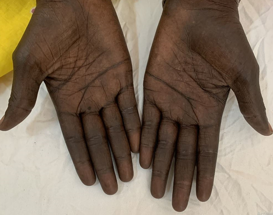
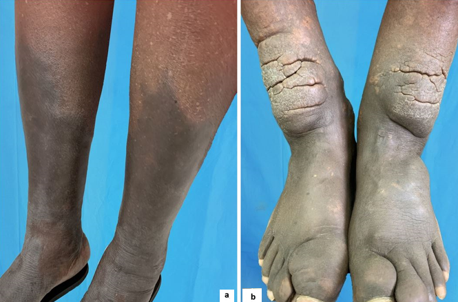
Among cutaneous signs ([Table 2]), pruritus, statistically correlated with the use of oral phytotherapy (p=0.04784) ([Table 3]), was found in 17% (n=36) of patients, and hyperpigmentation in 55.7% (n=117). The topography of hyperpigmentation ([Table 4]) was palmar ([Figure 1]) in 73.5% (n=86) of patients. In the 130 former patients, there was a statistically significant association between this hyperpigmentation and carbimazole (p=0.03721) and propranolol (p=0.009850) ([Table 3]). In all 188 patients, this hyperpigmentation was not statistically related to voluntary skin bleaching (p=0.5410) (Table III). Pretibial myxedema ([Figure 2]) was found in 1.4% of patients (n=3).
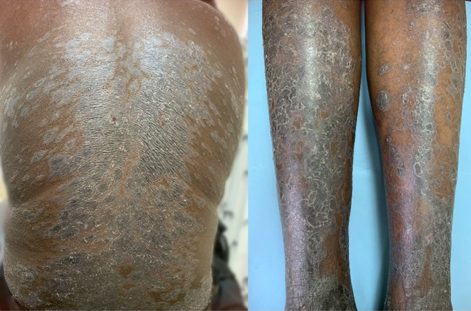
In relation to hair disorders (Table V), dry and brittle hair was reported in 47.6% (n=100) of patients. Dermatological manifestations demonstrated associations with inflammatory dermatosis in 16.6% (n=35), including acne in 12% (n=25) and superficial mycosis in 5.2% (n=11). Regarding nail disorders ([Table 4]), brittle nails were observed in 9% (n=19) of patients. Among former patients, lichenoid drug eruption ([Figure 3]) attributed to thiamazole was noted in one case.
Cardiovascular signs ([Table 7]) were present in 39.5% (n=83) of patients and were significantly associated with hyperpigmentation (p= 0.005752), hand wetness (p=0.04695), and pruritus (p=0.01346). Similarly, a statistically significant link was found between pruritus and digestive signs, present in 10% (n=21) of cases (p=0.01636).
Among patients with cutaneous manifestations, dysthyroidism evolution was observed in 68.6% (n=144), and of those, 70% (n=101) exhibited a favorable outcome with normalized thyroid hormone levels. Regarding the evolution of cutaneous manifestations in 105 patients, the condition remained stationary in 83.8% (n=88), comprising 68.2% (n=60) of old patients and 31.8% (n=28) of new patients. A positive evolution was noted in 16.2% (n=17), with 64.7% (n=11) being former patients and 35.3% (n=6) being new patients.


|
Auto-immune diseases |
Effective (n) |
Percentage (%) |
|
Type 1 diabetes |
2 |
0,9 |
|
Vitiligo |
3 |
1,4 |
|
Biermer's disease |
1 |
0,5 |
|
Systemic scleroderma |
1 |
0,5 |
|
Devic's neuromyelitis optica |
1 |
0,5 |
|
Skin disorders |
Old n (%) |
Recent n (%) |
Total (n) |
Frequency (%) |
|
Hyperpigmentation |
67 (51,5) |
50 (62,5) |
117 |
55,7 |
|
Xerosis |
31(23,8) |
8(10) |
39 |
18,6 |
|
Wetness of hands |
28 (21,5) |
20(25) |
48 |
22,8 |
|
Pruritus |
16(12,3) |
20(25) |
36 |
17 |
|
Palmoplantar keratoderma |
22(16,9) |
8(10) |
30 |
14,3 |
|
Hypersudation |
8(6) |
5(6,2) |
13 |
6,2 |
|
Palmar erythema |
0(0) |
4 (5) |
4 |
1,9 |
|
Melasma |
2(1,5) |
1(1,2) |
3 |
1,4 |
|
Pretibial myxedema |
3(2,3) |
0(0) |
3 |
1,4 |
|
Acropachydermia |
1(0,76) |
0(0) |
1 |
0,5 |
|
Chronic urticaria |
0(0) |
1(1,2) |
1 |
0,5 |
|
Medical treatment received |
Hyperpigmentation |
Without hyperpigmentation |
P-value |
|
|
Carbimazole |
Yes |
30 |
17 |
0.03721 |
|
No |
37 |
46 |
||
|
Thiamazole |
Yes |
35 |
42 |
0.09905 |
|
No |
32 |
21 |
||
|
Propranolol |
Yes |
33 |
17 |
0.009850 |
|
No |
34 |
46 |
||
|
Medical treatment received |
Alopecia |
Without alopecia |
|
|
|
Carbimazole |
Yes |
24 |
23 |
0.2199 |
|
No |
33 |
50 |
||
|
Thiamazole |
Yes |
29 |
48 |
0.09189 |
|
No |
28 |
25 |
||
|
Propranolol |
Yes |
25 |
25 |
0.2715 |
|
No |
32 |
48 |
||
|
Phytotherapy |
Pruritus |
Without pruritus |
|
|
|
Oral phytotherapy |
Yes |
9 |
20 |
0.04784 |
|
No |
27 |
154 |
||
|
Voluntary skin bleaching |
Hyperpigmentation |
Without hyperpigmentation |
|
|
|
Depigmentation |
Yes |
45 |
39 |
0.5410 |
|
No |
51 |
53 |
|
Hyperpigmentation Topography |
New (n) |
Old (n) |
Total (n) |
Frequency (%) |
|
Palmar |
36 |
50 |
86 |
73,5 |
|
Palmo - Plantar |
13 |
16 |
29 |
24,8 |
|
Upper limbs |
1 |
0 |
1 |
0,85 |
|
Generalized |
0 |
1 |
1 |
0,85 |
|
Total |
50 |
67 |
117 |
100 |
|
Skin involvement |
Old n (%) |
New n (%) |
Total (n) |
Frequency (%) |
|
Dry, brittle hair |
59(45,4) |
41(51) |
100 |
47,6 |
|
Alopecia |
57(43,8) |
38(47,5) |
95 |
45 |
|
Axillary depilation |
42 (32,3) |
34(42,5) |
76 |
36,2 |
|
Eyebrow tail sign |
20(15,4) |
14(17,5) |
34 |
16,2 |
|
Pubic depilation |
2(1,5) |
2(2,5) |
4 |
1,9 |
|
Nail disorders |
Old n (%) |
New n (%) |
Total n |
Frequency (%) |
|
Brittle nails |
11(8,5) |
8(10) |
19 |
9 |
|
Onychorrhexis |
5(3,8) |
1(1,2) |
6 |
2,9 |
|
Nail dystrophy |
1(0,8) |
1(1,2) |
2 |
0,9 |
|
Onycholysis |
2(1,5) |
0(0) |
2 |
0,9 |
|
Leuconychia |
1(0,8) |
0(0) |
1 |
0,5 |
|
Melanonychia |
1(0,8) |
0(0) |
1 |
0,5 |
|
Xanthonychia |
1(0,8) |
0(0) |
1 |
0,5 |
|
Extra-dermatological signs |
Hyperpigmentation |
Without hyperpigmentation |
P-value |
|
|
Cardiovascular |
Yes |
56 |
27 |
0.005752 |
|
No |
61 |
66 |
||
|
Neuropsychiatric |
Yes |
40 |
21 |
0.06754 |
|
No |
77 |
72 |
||
|
Osteoarticular |
Yes |
14 |
5 |
0.1035 |
|
No |
103 |
88 |
||
|
|
|
Pruritus |
Without pruritus |
|
|
Cardiovascular |
Yes |
21 |
62 |
0.01346 |
|
No |
15 |
112 |
||
|
Digestive |
Yes |
8 |
13 |
0.01636 |
|
No |
28 |
161 |
||
|
|
|
Wetness of hands |
Without wetness of hands |
|
|
Exophthalmos |
Yes |
38 |
95 |
0.008746 |
|
No |
10 |
67 |
||
|
Cardiovascular |
Yes |
25 |
58 |
0.04695 |
|
No |
23 |
104 |
Discussion
This cross-sectional study presents findings from prospective data collection conducted over a 6-month period, involving 288 patients diagnosed with Graves' disease, of whom 210 exhibited dermatological manifestations, indicating a prevalence of 72.9%.
Despite the widespread impact of Graves' disease, there remains a scarcity of studies in Africa addressing dermatological manifestations in this context. In a Tunisian study by Mansour et al,[5] which included 32 patients with hyperthyroidism, all participants presented with dermatological manifestations. Conversely, in Asia, a Malaysian study by Ramanathan et al[4] involving 236 patients with hyperthyroidism reported a prevalence of 18.75% with one or more dermatological manifestations. Similarly, an Indian study by Bains et al [6], spanning a 1-year period and encompassing 13 patients with hyperthyroidism, documented dermatological manifestations in 69.23% of cases.
Pruritus is a recognized but uncommon characteristic sign in Graves' disease.[7] In our study, it was identified in 17% (n=36) of patients presenting dermatological signs, constituting 12.5% (36/288) of all Graves' disease cases. A Malaysian study by Ramanathan et al[4] reported pruritus in 6.4% of patients,[4] while an American study by Caravati et al, [8] involving 154 hyperthyroidism cases, found pruritus in 5.8%, particularly in cases of untreated, long-term Graves' disease. Mansour et al[5] in Tunisia reported a higher incidence of pruritus, reaching 31% in their patient cohort. According to some authors, this pruritus may result from the activation of kininogens to kinins by kallikreins, leading to increased tissue metabolism.[4], [9] In our patients, the frequency of pruritus appeared to be exacerbated by the use of medicinal plants, showing a statistically significant correlation with this symptom (p=0.04784). The literature associates various medicinal plants with pruritus,[10] including Anacardium occidentale.[11] implicated in plant-induced dermatoses in Senegal.[12] Consequently, persistent pruritus in a well-controlled hyperthyroid patient warrants investigation, and caution should be exercised with herbal remedies.
Hyperpigmentation, observed in 55.7% (n=117) of patients with dermatological manifestations, manifested in 40.6% (117/288) of all Graves' disease cases. These findings align with Anania et al[13] series of 32 patients (38%) and Srujana et al[14] series of 43 patients (40%). However, they are lower than Michelangeli et al[15] reported incidence of 73% in 194 patients with Graves' disease in Papua New Guinea. The prevailing hypothesis suggests increased pituitary ACTH release compensating for accelerated cortisol degeneration, contributing to hyperpigmentation through heightened melanotropic activity, rather than hemosiderin deposition due to capillary fragility.[16], [17] Literature often describes hyperpigmentation in Graves' disease as localized to the face or diffuse,[15] with greater prominence in individuals with a darker phototype.[18] Despite this, the correlation between carbimazole and skin hyperpigmentation remains paradoxical. While its metabolite, methimazole (thiamazole), has been implicated in skin hyperpigmentation, studies on melanocyte cultures indicate a depigmenting effect by inhibiting melanin synthesis. [19] This action is achieved through the inhibition of peroxidase in cutaneous melanocytes, affecting the metabolism of melanin intermediates, such as dihydroxyphenylalanine (DOPA), dihydroxyindole, and benzothiazine.[20], [21], [22] Carbimazole's most frequent cutaneous side effects in the literature are maculopapular exanthema and urticaria, with rare cases of exfoliative dermatitis and lupus erythematosus-like syndrome reported.[23] A case of lichenoid toxidermia to carbimazole or its metabolite (thiamazole) is infrequently reported, as observed in one of our patients.
Furthermore, propranolol, statistically linked to skin hyperpigmentation (p=0.002), is recognized as a potential contributor to this dermatological sign.[19] Tatu et al[24] reported that beta-blockers, including propranolol, can induce skin hyperpigmentation, particularly of the lichenoid type.
On the basis of this observation, the persistent nature of cutaneous hyperpigmentation during hyperthyroidism, despite the effectiveness of antithyroid treatment, may be attributed to the specific therapeutic agents employed. Furthermore, the statistically significant associations between hyperpigmentation and osteoarticular (p=0.017), cardiovascular (p=0.001), and neuropsychiatric (p=0.025) disorders underscore the importance of considering cutaneous hyperpigmentation as a potential indicator of severity in patients with hyperthyroidism.
Contrary to the various manifestations discussed, pretibial myxedema was identified in only 1.04% (n=3) of our patients. Similar findings were observed in the studies of Ramanathan et al[4] in Malaysia and Kee et al[25] in Singapore, reporting frequencies of 1.3% (n=3) and 0.7%, respectively. However, these figures are lower than those reported by Razi et al[26] in Pakistan, who documented a frequency of 6.5% (n=8) of pretibial myxedema in their series. These consistent findings reinforce the rarity of pretibial myxedema in hyperthyroidism. This particular manifestation arises from the excessive synthesis of glycosaminoglycans by fibroblasts, prompted by the binding of TSH receptor autoantibodies to receptors on the surface of these cells.[27] The consequence is an accumulation of mucin in the dermis and hypodermis, occasionally leading to compression of dermal lymphatic vessels.[28]
Within the subset of cases presenting with inflammatory dermatosis, accounting for 12% (n=35) of the cohort, acne stood out as the primary manifestation, manifesting in 9% of cases. Existing literature highlights an association between acne and autoimmune thyroiditis. Furthermore, dysthyroidism is implicated in the severity of acne, irrespective of the specific type of thyroid dysfunction.[29], [30]
Conclusion
Frequent dermatological manifestations characterize Graves' disease, with a predominant feature being cutaneous hyperpigmentation, particularly evident in the palmoplantar region. However, the administration of treatments may influence the occurrence and persistence of these manifestations.
Source of Funding
None.
Conflict of Interest
None.
References
- JD Safer. Thyroid hormone action on skin. Curr Opin Endocrinol Diabetes Obes 2012. [Google Scholar]
- E Acer, E Ağaoğlu, G Yorulmaz, HK Erdoğan, ES Alagüney, ZN Saraçoğlu. Evaluation of cutaneous manifestations in patients with thyroid diseases under treatment. Turkderm - Turk Arch Dermatol Venereol 2020. [Google Scholar]
- JB Sindhu, KR Reddy, GNR Netha, DS Vani. Study of cutaneous manifestations in thyroid disorders. J Evolution Med Dent Sci 2016. [Google Scholar]
- M Ramanathan, MN Abidin, M Muthukumarappan. The prevalence of skin manifestations in thyrotoxicosis--a retrospective study. Med J Malaysia 1989. [Google Scholar]
- Y Mansour, A Saoussi, CB Slama, H Slimane, M. Mokni. Manifestations cutanées des dysthyroïdies. Ann Dermatol Vénéréologie 2018. [Google Scholar]
- A Bains, GR Tegta, V Deepak. A cross-sectional study of cutaneous changes in patients with acquired thyroid disorders. Clin Dermatol Rev 2019. [Google Scholar]
- C Tan, K Loh. Generalised pruritus as a presentation of Grave's disease. Malays Fam Physician 2013. [Google Scholar]
- CM Caravati Jr, DR Richardson, BT Wood. Pruritus and excoriations in hyperthyroidism. South Med J 1969. [Google Scholar]
- M Krajnik, Z Zylicz. Understanding Pruritus in Systemic Disease. J Pain Symptom Manage 2001. [Google Scholar]
- S T Shimray, H K Sharma. A review on:Itch-causing and itch-relieving plants. Curr Trends Pharm Res 2022. [Google Scholar]
- MO Hamburger, GA Cordell, N Ruangrungsi. Traditional medicinal plants of Thailand XVII Biologically active constituents of Plumeria rubra. J Ethnopharmacol 1991. [Google Scholar]
- SO Niang, BA Diatta, Y Tine, S Diadié, K Diop, C Ndiaye. Prise en charge des hypersensibilités retardées aux plantes médicinales. Rev Fr Allergol 2020. [Google Scholar]
- C Anania, S Rubenfeld. Hyperpigmentation in Graves' disease. Thyroidology 1989. [Google Scholar]
- B Srujana, BN Reddy, G Prasad. Clinical spectrum of cutaneous manifestations of thyroid disorders in patients attending MediCiti Institute of Medical Sciences. IP Indian J Clin Exp Dermatol 2016. [Google Scholar]
- VP Michelangeli, G Pawape, A Sinha, K Ongugu, D Linge, SH Sengupta. Clinical features and pathogenesis of thyrotoxicosis in adult Melanesians in Papua New Guinea. Clin Endocrinol (Oxf) 2000. [Google Scholar]
- JM Leonhardt, WR Heymann. Thyroid disease and the skin. Dermatol Clin 2002. [Google Scholar]
- X Song, Y Shen, Y Zhou, Q Lou, L Han, J K Ho. General hyperpigmentation induced by Grave's disease: A case report. Medicine (Baltimore) 2018. [Google Scholar] [Crossref]
- R Miller, FS Ashkar, J Jacobi. Hyperpigmentation in Thyrotoxicosis. JAMA 1970. [Google Scholar] [Crossref]
- RM Giménez García, SC Molina. Drug-Induced Hyperpigmentation: Review and Case Series. J Am Board Fam Med 2019. [Google Scholar]
- B Kasraee, A Hugin, C Tran, O Sorg, JH Saurat. Methimazole is an inhibitor of melanin synthesis in cultured B16 melanocytes. J Invest Dermatol 2004. [Google Scholar]
- B Kasraee. Peroxidase-mediated mechanisms are involved in the melanocytotoxic and melanogenesis-inhibiting effects of chemical agents. Dermatology 2002. [Google Scholar]
- B Kasraee. Depigmentation of brown Guinea pig skin by topical application of methimazole. J Invest Dermatol 2002. [Google Scholar]
- GA Brent, RJ Koenig, LL Brunton, BA Chabner, BC Knollmann. Thyroid and anti-thyroid drugs. Goodman & Gilman‟s pharmacological basis of therapeutics 2011. [Google Scholar]
- AL Tatu, AM Elisei, V Chioncel, M Miulescu, LC Nwabudike. Immunologic adverse reactions of β-blockers and the skin. Exp Ther Med 2019. [Google Scholar]
- CW Kee, JS Cheah. Thyrotoxic pretibial myxoedema in Asian patients in Singapore. Postgrad Med J 1975. [Google Scholar]
- A Razi, F Golforoushan, AB Nejad, M Goldust. Evaluation of dermal symptoms in hypothyroidism and hyperthyroidism. Pak J Biol Sci 2013. [Google Scholar]
- F Cianfarani, E Baldini, A Cavalli, E Marchioni, L Lembo, M Teson. TSH Receptor and Thyroid-Specific Gene Expression in Human Skin. J Invest Dermatol 2010. [Google Scholar]
- R H Bull, P S Mortimer, P R Coburn. Pretibial myxoedema: a manifestation of lymphoedema?. Lancet 1993. [Google Scholar]
- L Endres, DM Tit, S Bungau, NA Pascalau, LM Todan, E Bimbo-Szuhai. Incidence and Clinical Implications of Autoimmune Thyroiditis in the Development of Acne in Young Patients. Diagnostics (Basel) 2021. [Google Scholar] [Crossref]
- T Vergou, E Mantzou, P Tseke, A E Moustou, A Katsambas, M Alevizaki. Association of thyroid autoimmunity with acne in adult women. J Eur Acad Dermatol Venereol 2012. [Google Scholar]
